
Explainer · August 5, 2026 · 4 min · By Noor El-Amin
Ultrasound Before the Syringe: Why Beverly Hills Injectors Are Mapping Faces First
High-frequency ultrasound is quietly changing how filler is placed, dissolved, and corrected in one of the busiest injectable markets in the country. Here is what the technology actually does, what it cannot do, and how patients can ask about it.
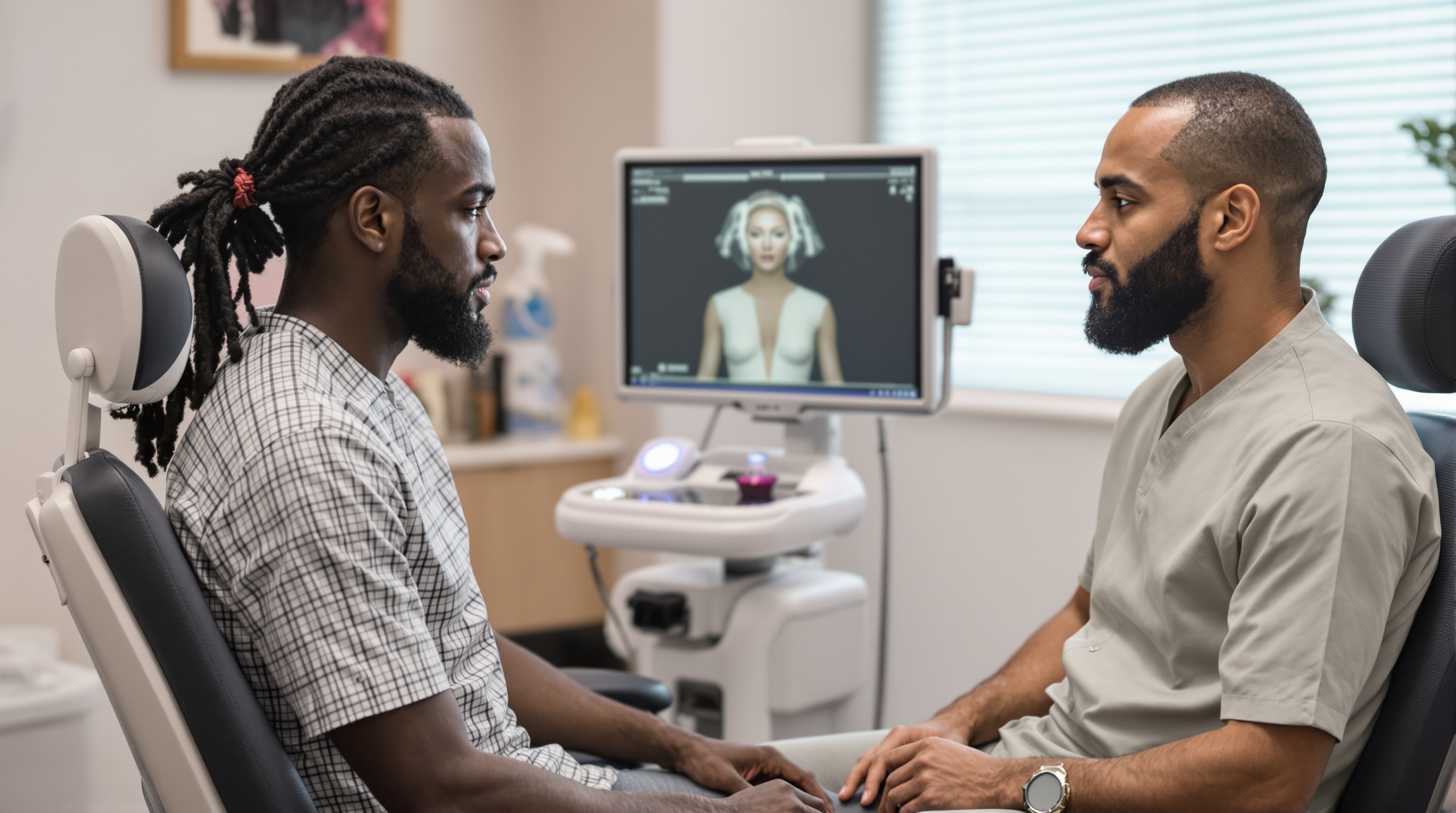
Walk into a high-volume aesthetic practice in Beverly Hills today and there is a growing chance the first device that touches your face is not a needle. It is an ultrasound probe. Point-of-care ultrasound, often shortened to POCUS, has moved from emergency rooms and vascular labs into dermatology suites, and it is reshaping how hyaluronic acid filler is planned, placed, and reversed.
What the probe actually shows. High-frequency ultrasound, typically in the 15 to 24 megahertz range for facial work, produces a real-time image of the skin, subcutaneous fat, muscle, and vessels down to roughly two to three centimeters of depth. Hyaluronic acid filler appears as a dark, well-defined pocket. Arteries show pulsatile flow on Doppler mode. That combination matters for two reasons. First, injectors can see where old product from previous appointments actually sits, which is often not where the patient or the prior injector believes it sits. Second, they can visualize the facial artery, the angular artery, and other vessels whose paths vary meaningfully from person to person. Cadaver studies have repeatedly shown that so-called standard vascular anatomy is a statistical average, not a guarantee.
Why this matters in a filler-saturated market. Beverly Hills has an unusually high density of patients with long injection histories, sometimes spanning fifteen or twenty years and multiple providers. Layered, migrated, or partially degraded filler is common. When a new injector adds product on top of unmapped older material, the risks include lumpiness, delayed nodules, and the puffy, overfilled look that patients increasingly want to avoid. Ultrasound lets a clinician answer a basic question before injecting anything: what is already in this face, and where?
The dissolving problem. The same logic applies to hyaluronidase, the enzyme used to dissolve hyaluronic acid filler. Blind dissolving, meaning injecting the enzyme into the general area where filler is suspected, often requires multiple sessions and higher total doses, because the enzyme only works where it physically contacts product. Ultrasound-guided hyaluronidase allows the clinician to place the enzyme directly into the visualized filler pocket. Published case series suggest this targeted approach can reduce both the number of sessions and the total enzyme dose, which matters because hyaluronidase can also degrade some native hyaluronic acid and carries a small allergy risk.
The emergency use case. The most serious filler complication is vascular occlusion, where product enters or compresses an artery and cuts off blood supply to skin, or in rare cases, to the retina. Time matters enormously here. Ultrasound can help confirm reduced or absent flow in a suspected vessel and guide hyaluronidase precisely to the site of obstruction rather than flooding the entire region. It does not replace clinical judgment, and treatment should never be delayed to hunt for imaging, but as an adjunct it can make a rescue protocol more targeted.
What ultrasound cannot do. A few honest limits. Ultrasound cannot reliably identify which brand of filler is present, and it struggles to distinguish some biostimulatory products and older permanent fillers from scar tissue without an experienced eye. Image quality is heavily operator dependent: a probe in untrained hands produces confident-looking pictures that mean very little. Formal training in facial sonoanatomy takes real time, and there is currently no universal certification standard specific to aesthetic ultrasound in the United States. It also adds cost and appointment length, which is why many practices reserve it for revision cases, dissolving sessions, high-risk zones like the nose and glabella, and complication workups rather than every routine syringe.
Questions worth asking. Patients considering filler, especially anyone with prior filler from another provider, can reasonably ask three things. One: do you use ultrasound, and in which situations? Two: if I need dissolving, is it done under guidance or blind? Three: what is your protocol if a vascular event happens during my appointment? A practice does not need to image every patient to be safe, but it should have clear, confident answers.
The bottom line. Ultrasound guidance does not make filler risk-free, and it is not a substitute for deep anatomical knowledge. What it does is convert assumptions into observations. In a market where faces often carry years of accumulated product, seeing before injecting is a meaningful step forward, and its spread through Beverly Hills practices reflects a broader shift in aesthetics: away from volume as the goal, toward precision as the standard.
Related reading: Hyaluronic Acid Fillers vs. Biostimulators: What Beverly Hills Injectors Are Actually Choosing, and Why.
More in Explainer
View all →- RF Microneedling vs. Traditional Microneedling: What the Energy Actually Adds
- Why Melasma Often Gets Worse After Laser, and What Actually Works
- Fillers That Fill vs. Fillers That Build: How Biostimulatory Injectables Actually Work
- Sculptra vs. Radiesse: How Beverly Hills Injectors Actually Choose Between the Two Biostimulators